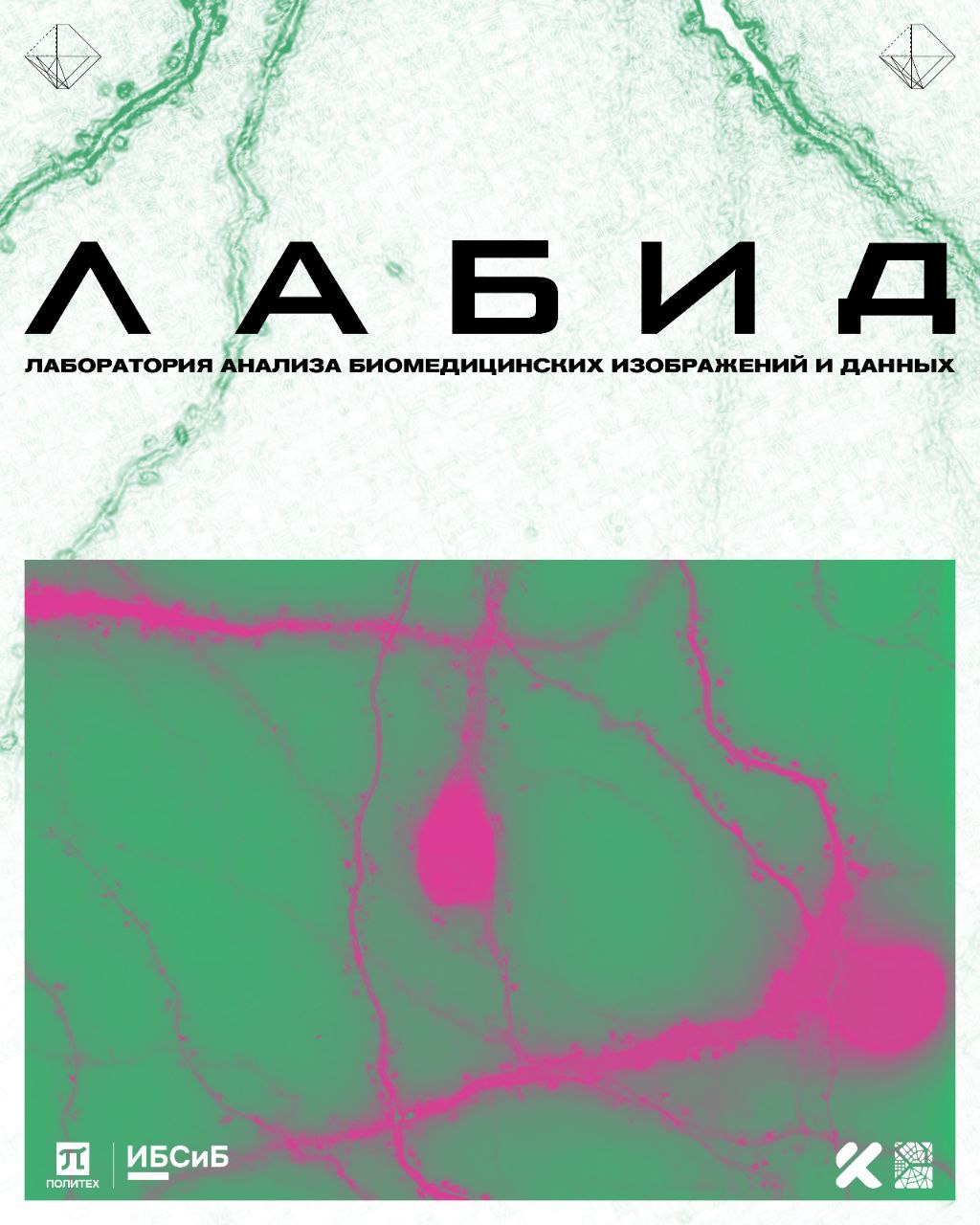
3D image processed by a neural network
2048×2048×32 on RTX 3080Biomedical Imaging & Data Analysis Lab · SPbPU
Exploring biomedical data with artificial intelligence
We develop algorithms and software tools for microscopy, neuron analysis and scientific data — from fundamental neuroscience to reproducible digital methods.
01 biomedical data × artificial intelligence
Method results
Measurable improvements in microscopy data processing
PSNR of neural-network deconvolution
vs. 31.2 dB for the Richardson–Lucy methodRMSE reduction
for the developed denoising methodPSNR increase
compared with traditional denoising methodsResearch
At the intersection of neuroscience, computer vision and data engineering
The laboratory brings together biologists, programmers and AI researchers. Work focuses on practical tools that help extract more information from complex biomedical data.
Microscopy images
Denoising, deconvolution and quality enhancement of 3D fluorescence images with neural-network and classical methods.
Neuron morphology
Segmentation, measurement and classification of dendritic spines to study synaptic plasticity and neurodegenerative changes.
Neuronal activity
Analysis of activity time series of individual neurons and neural networks, including interpretable machine-learning models.
Scientific data and knowledge
Tools for annotating, reusing and searching research data, plus retrieval-augmented generation systems for biomedical tasks.
Key projects
Tools for biomedical data analysis
Image restoration
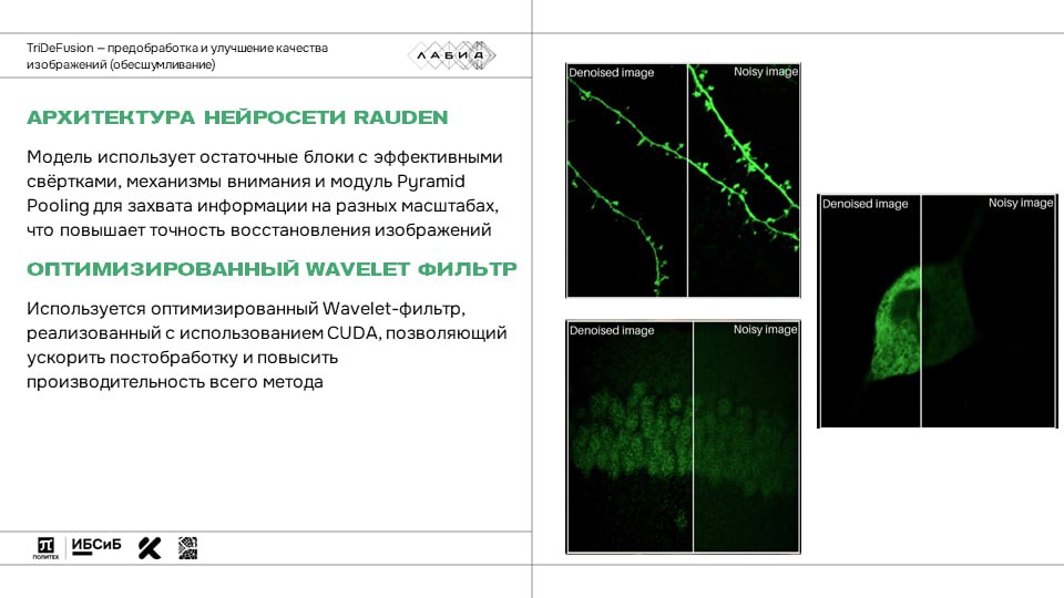
TriDeFusion + FRT
A hybrid denoising method and a cloud service for fluorescence microscopy data.
TriDeFusion combines RAUDen neural-network denoising with optimized wavelet filtering. FRT moves processing to the cloud, letting users run denoising and deconvolution without their own computing resources.
- 3D fluorescence microscopy
- RAUDen + Wavelet
- cloud processing
Deep learning
NeuroDecon
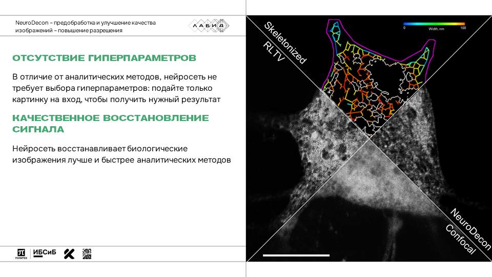
A neural-network algorithm for super-resolution of 3D microscopy images.
The method uses calibration fluorescence microspheres and mathematically simulated objects to generate training data. The architecture combines U-Net and residual blocks, making volumetric image processing more efficient.
- U-Net
- residual blocks
- 3D deconvolution
Publications International Journal of Molecular Sciences, 2025
Code NeuroDecon
Neuron morphology
SpineTool
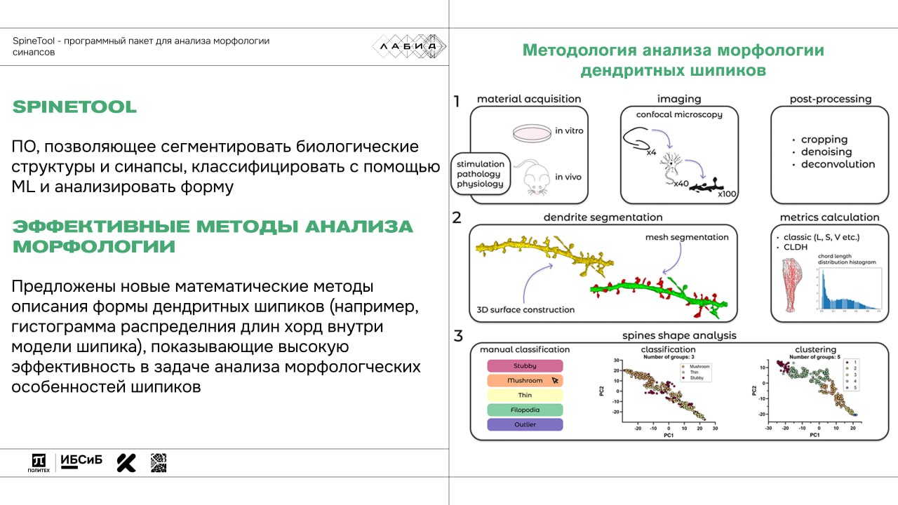
Open-source software for comprehensive analysis of dendritic spines.
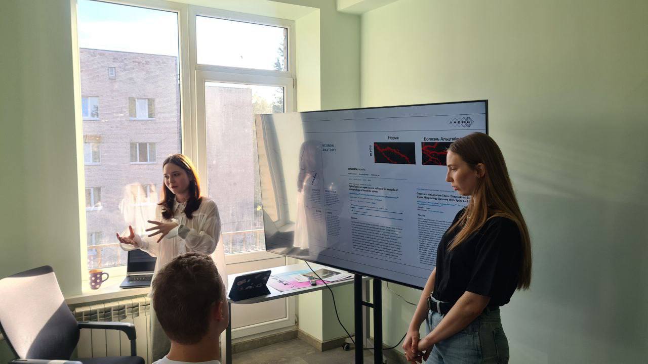
SpineTool combines semi-automatic segmentation, morphological parameter calculation, clustering, manual labeling and classification of dendritic spines. The tool is used to study synaptic plasticity and Alzheimer's disease models.
- 3D segmentation
- morphometry
- classification
Code SpineTool
Neuronal activity
NeuroActivityToolkit
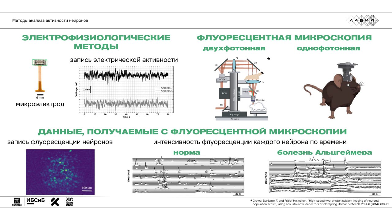
A toolkit for quantitative analysis of fluorescence recordings of neuronal activity.
The project automates the calculation of statistical characteristics of activity time series of individual neurons and the neuronal network. The approach is used to compare activity and study effects related to neurodegenerative diseases.
- calcium imaging
- time series
- neural networks
Publications Journal of Imaging, 2023
Scientific search
NeuroRAG
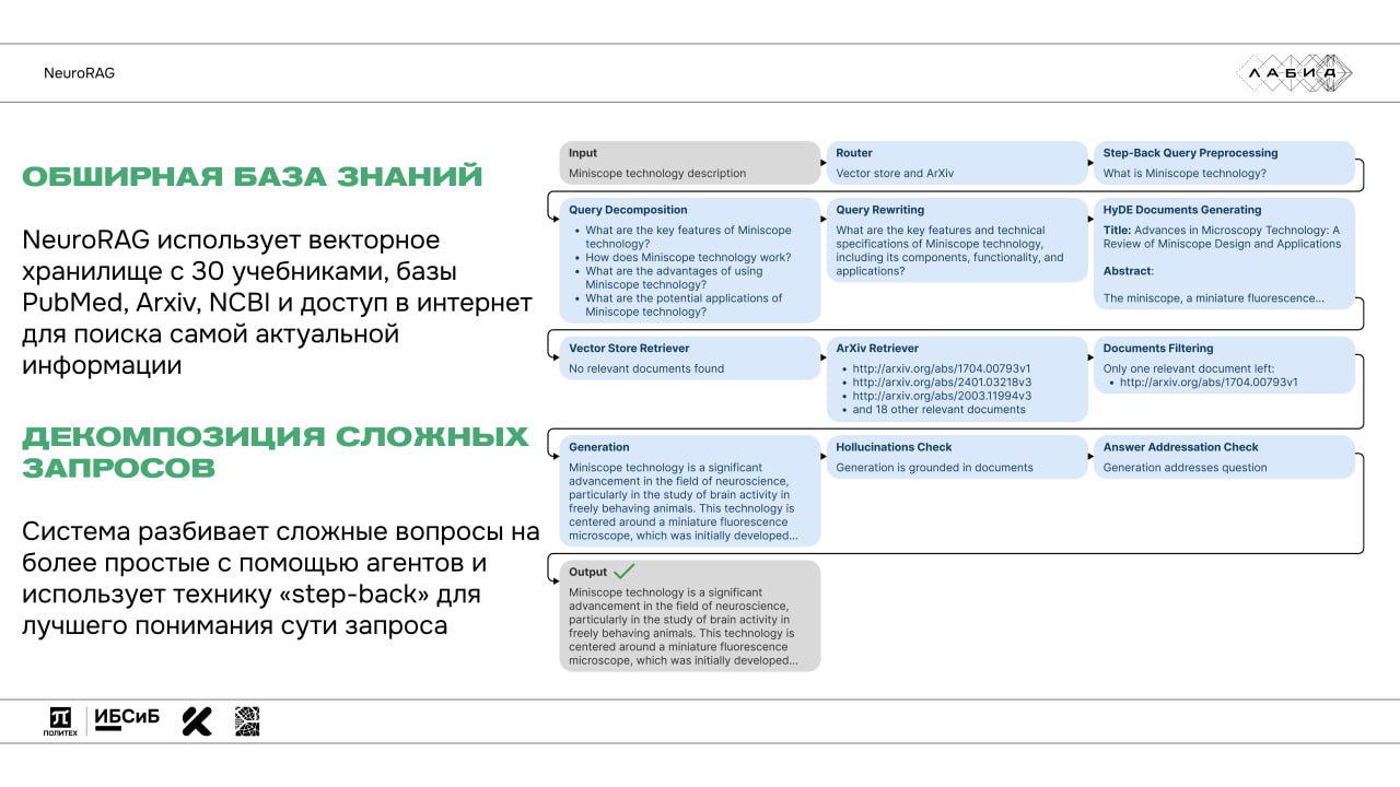
A multi-agent question-answering system for neuroscience.
NeuroRAG combines large language models and retrieval-augmented generation: it decomposes complex questions, retrieves data from domain sources and applies extra filtering and verification steps to the answers.
- RAG
- multi-agent
- scientific sources
Code NeuroRAG
Explainable AI
NEuRT
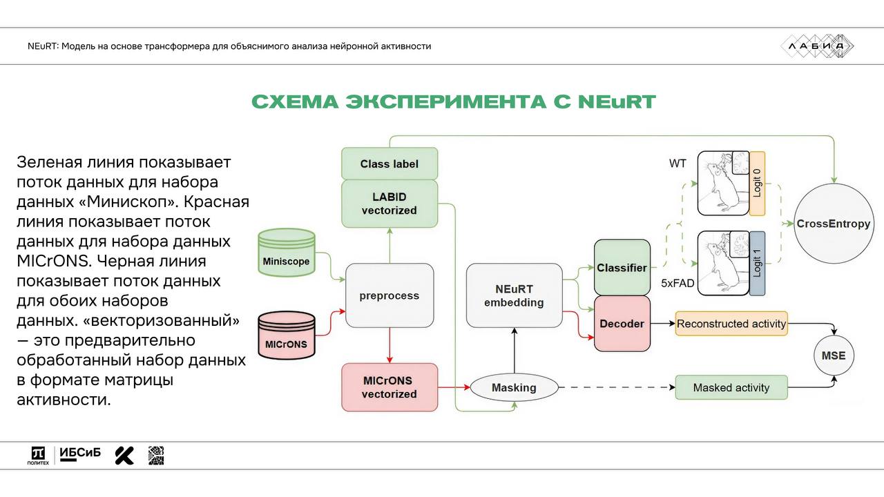
A transformer model for analyzing and interpreting the activity of real brain networks.
NEuRT is pretrained on the open MICrONS dataset and adapts to small user samples. Interpretation methods show which activity patterns drive the model's decisions.
- Transformer
- Attention Rollout
- MICrONS ~270 GB
Laboratory
An environment where biology and software engineering work together
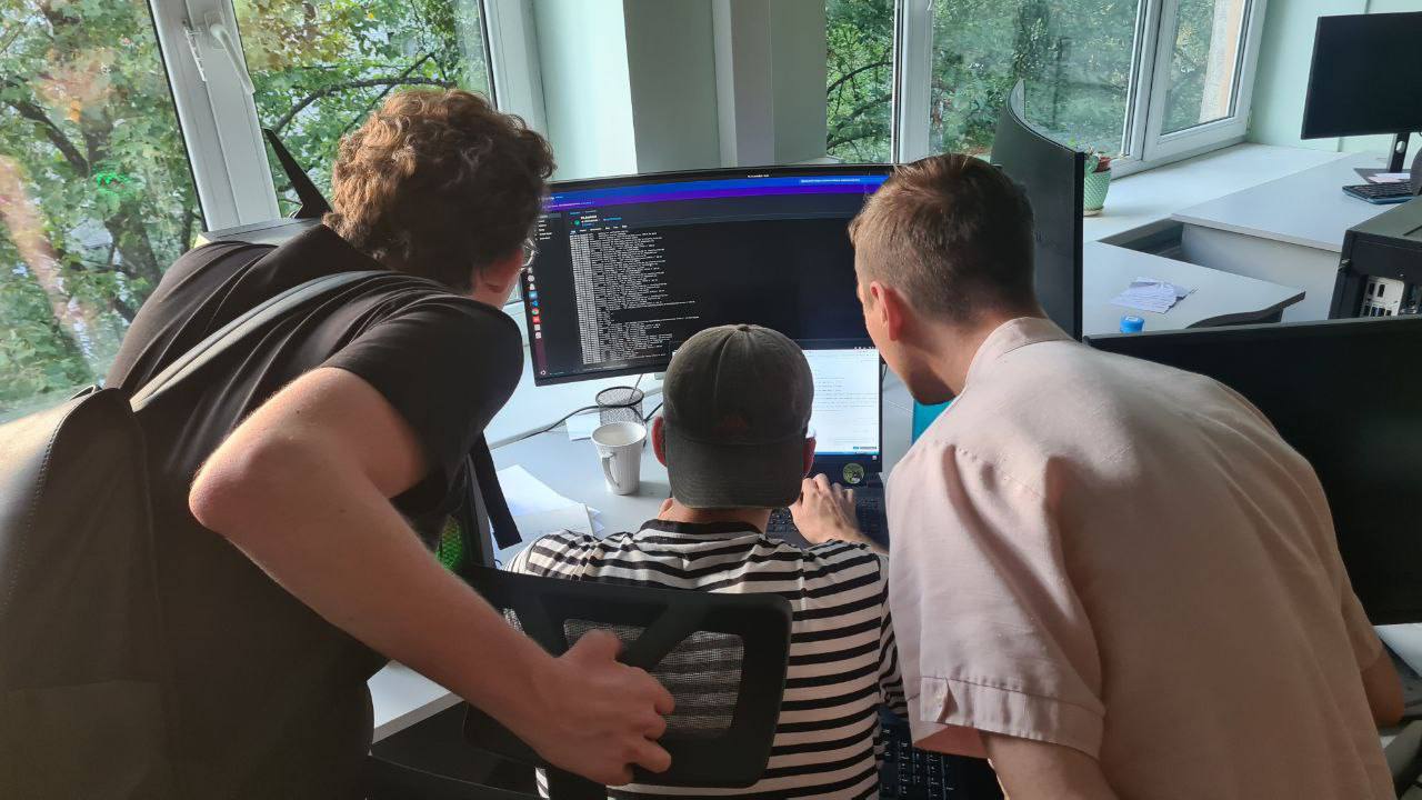
LABID was founded at Peter the Great St. Petersburg Polytechnic University. The team unites researchers of different backgrounds and takes projects from a biological question to software implementation and a scientific publication.
Contacts
Open to scientific collaboration
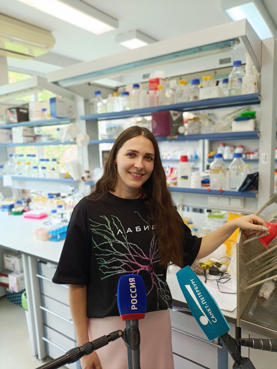
Head of the laboratory
Ekaterina I. Pchitskaya, PhD
Associate professor at the Higher School of Biomedical Systems and Technologies, SPbPU. Research at the intersection of neuroscience, microscopy image analysis and artificial intelligence.